WITH THE SINUS, CHECK THE JAW: EDITION FIVE
When Chronic Sinusitis Is a Tooth Problem That No One Has Found Yet
By Dr. Frits Hoogendijk | Maxillofacial Surgeon | Menlopark Medical Centre, Pretoria | Benoni
Contact: 082 390 7425
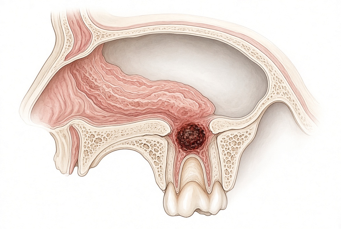
In Edition Four of this series, we established that ear pain should always prompt a concurrent assessment of the jaw — because up to half of all otalgia in adults originates not in the ear, but in the structures it shares its nerve supply with, including the teeth and jaw. Normal otoscopy is not the end of the diagnostic process. It is the beginning of the question: if not the ear, where?
Edition Five addresses a parallel scenario that is, if anything, even more frequently encountered in clinical practice, and even more frequently mismanaged: the patient with chronic or recurrent maxillary sinusitis that does not respond to medical treatment, does not resolve with antibiotics, and returns repeatedly despite ENT consultation and sinus CT scanning — because the sinus is not the origin of the problem. The tooth is.
With the sinus, check the jaw.
Odontogenic Sinusitis: The Diagnosis That Is Hiding in Plain Sight
The maxillary sinus sits in intimate proximity to the roots of the upper posterior teeth. The roots of the upper first and second molars — and frequently the upper second premolar — project directly into or immediately beneath the floor of the maxillary sinus, separated in many individuals by only a thin layer of cortical bone or, in some cases, by no bone at all. In a significant proportion of the population, the root apices of the upper molars are separated from the sinus cavity by less than one millimetre of tissue.
This anatomical relationship creates a direct pathway for dental infection to invade the sinus. When a periapical granuloma, a periapical cyst, or a chronic endodontic infection develops at the apex of an upper molar, the inflammatory process does not respect the sinus floor. Over time, it erodes through — directly seeding the sinus mucosa with bacteria and inflammatory mediators from the dental source. The result is sinusitis. But the source is the tooth, not the sinus.
This condition — odontogenic maxillary sinusitis (OMS) — is extraordinarily common, yet consistently underdiagnosed. Approximately one quarter of all chronic maxillary sinusitis cases are odontogenic in origin. When only unilateral maxillary sinusitis is considered — as is characteristic of dental origin, since teeth are unilateral structures — the proportion rises dramatically: 40% of unilateral maxillary sinusitis is odontogenic. In studies where ipsilateral dental pathology was simultaneously present, the prevalence of OMS reached 79.63%.
These figures are not marginal. They mean that in a patient presenting with chronic sinusitis on one side, there is a near-one-in-two chance that the source is a tooth — and that no amount of nasal decongestant, steroid spray, or antibiotic therapy will resolve it, because the infection is being continuously replenished from the dental source.
A Word on Bilaterality: Not as Reassuring as It Seems
The conventional teaching is that odontogenic sinusitis is characteristically unilateral — and this is true in the majority of cases. However, clinicians should be aware of an important caveat that is consistently overlooked: bilateral odontogenic sinusitis does occur.
In our maxillofacial surgery practice, we see patients presenting with bilateral maxillary sinusitis who are assumed to have primary inflammatory or allergic sinus disease — because bilateral pathology is considered inconsistent with a dental origin. This assumption is not always correct.
Patients with multiple heavily restored upper molar and premolar teeth — patients who have had extensive dental treatment over many years, including multiple root canal treatments, crowns, and large restorations — may have bilateral upper posterior dental pathology simultaneously. A failed root canal on the upper right first molar and a periapical granuloma on the upper left second molar, both eroding into their respective sinus floors, will produce bilateral maxillary sinusitis. The presentation may appear indistinguishable from primary bilateral sinusitis on medical CT scanning — because, as discussed below, medical CT misses the dental source in the majority of cases regardless of laterality.
Bilateral OMS is not common. But it occurs with sufficient regularity in patients with a history of extensive upper posterior dental work that bilateral sinusitis should not automatically be used to exclude the jaw from the diagnostic process. In any patient with chronic bilateral maxillary sinusitis and a history of multiple upper molar restorations, root treatments, or crowns, CBCT assessment of the upper posterior dentition bilaterally is warranted.
The rule remains: with the sinus, check the jaw — even when both sinuses are involved.
Why the Sinus CT and the Dental X-Ray Both Miss the Diagnosis
The diagnostic failure in odontogenic sinusitis is not the result of inadequate clinical care. It is the result of a systematic mismatch between the imaging tools used and the pathology being sought.
When a patient presents with chronic sinusitis, the standard imaging investigation is a medical CT scan of the sinuses. This is the correct tool for demonstrating mucosal thickening, sinus opacification, polyp formation, and anatomical variants relevant to surgical planning. It is an excellent tool for confirming sinusitis as a diagnosis. It is a poor tool for identifying the tooth that is causing it.
The reason is resolution and calibration. Medical CT of the sinuses is optimised for soft tissue and bony sinus architecture at a resolution appropriate for demonstrating centimetre-scale pathology. The periapical lesions responsible for odontogenic sinusitis are frequently millimetre-scale — a granuloma of 2–4mm at the root apex, a thin linear erosion through the sinus floor, a subtle discontinuity in the cortical plate. Medical CT, reconstructed in planes designed for sinus assessment, consistently misses these fine dental details.
The published evidence is stark. In two separate case series evaluating odontogenic sinusitis, approximately two-thirds of all identifiable dental pathology went unreported by radiologists reviewing sinus CT scans. These were not inadequate radiologists. They were reading the CT correctly — the dental pathology was simply below the detection threshold of the imaging modality and reporting framework they were using.
The failure on the dental side is equally consistent. Routine dental examination using periapical radiographs failed to diagnose odontogenic sinusitis in 86% of cases. The physical limitation is the same as described throughout this series: conventional 2D dental radiography requires substantial cortical bone destruction before a periapical lesion becomes visible. Small lesions that have already eroded into the sinus floor remain radiographically invisible on periapical films and OPG.
The result is a diagnostic gap that sits precisely between two specialties. The ENT specialist's CT confirms sinusitis but cannot resolve the dental source. The dentist's panoramic X-ray shows no obvious pathology. Neither investigation has used the right tool for the question that matters: is there a dental lesion at the root apex of the upper molars eroding into the sinus floor?
What CBCT Sees That Everything Else Misses
Cone beam CT is calibrated specifically for the tooth-bone-sinus interface — the precise anatomical region where odontogenic sinusitis originates. Its voxel resolution of less than half a millimetre, combined with multiplanar reconstruction in the dental axes, makes it the optimal tool for visualising the relationship between the root apex, the sinus floor cortex, and the sinus mucosa in three dimensions.
The diagnostic performance data are compelling:
- CBCT detected 34% more periapical lesions than periapical radiography in posterior maxillary teeth, including significantly more lesions with expansion into the maxillary sinus
- CBCT identified mucosal changes associated with dental infections with a prevalence of 77%, compared to only 19% using conventional radiographs in the same patients
- CBCT identified sinus membrane thickening more than four times as often as periapical radiographs
- Periapical radiography identified only 3% of all apical infections extending into the sinus that were visible on CBCT
- CBCT has higher resolution than medical CT for identifying odontogenic lesions and their relationship with the maxillary sinus floor, and is now widely used as the preferred modality for OMS diagnosis
- CBCT carries approximately 10% lower radiation dose than medical CT and shorter examination time
The expert consensus on odontogenic maxillary sinusitis is explicit: while medical CT can identify sinus disease clearly, many dental lesions are difficult to identify on CT in detail, and CBCT — with its higher sensitivity for odontogenic lesions and the tooth-sinus floor relationship — is now the preferred diagnostic tool for OMS.
The Characteristic That Should Trigger the Dental Diagnosis: Unilaterality
There is a clinical sign in the presentation of odontogenic sinusitis that is both diagnostically powerful and consistently underutilised: unilaterality.
Primary sinusitis — viral, allergic, bacterial — typically involves both sinuses or presents with bilateral symptoms. Teeth, by contrast, are unilateral structures. A periapical infection at the root of the upper right first molar will drive inflammation into the right maxillary sinus only. Odontogenic sinusitis is therefore characteristically unilateral — affecting one maxillary sinus, on the same side as the causative tooth, with the contralateral sinus normal or minimally involved.
In a patient presenting with unilateral maxillary sinus opacification, this single finding should immediately elevate the probability of a dental origin. The expert consensus recommends that otorhinolaryngologists direct patients with unilateral maxillary sinus opacification to dental experts for evaluation of potential dental pathology — even in cases where no dental abnormalities are detected on their CT scans.
This is the key clinical instruction: a normal sinus CT does not exclude a dental source. The resolution is wrong for the question.
As noted above, bilaterality should not reflexively exclude the dental hypothesis in patients with extensive upper posterior dental work — bilateral dental pathology producing bilateral OMS is uncommon but real.
The Absent Toothache — Again
The pattern is identical to that observed in referred otalgia, TMJ dysfunction, and trigeminal neuralgia misdiagnosis throughout this series: the causative tooth almost never hurts.
The periapical granuloma eroding into the sinus floor is typically the product of longstanding chronic apical periodontitis — a condition that, as established in previous editions, is asymptomatic in the majority of patients. The patient has no toothache. They have sinusitis. They do not connect the two because there is nothing connecting them in their conscious experience. The tooth is silent. The sinus is inflamed.
When asked whether their teeth hurt, the patient answers no. The dental hypothesis is abandoned. The GP prescribes more antibiotics. The ENT performs functional endoscopic sinus surgery. The sinus improves temporarily. The tooth continues to seed the sinus. The sinusitis returns.
The dental source was never found — because no one used the right imaging tool to look for it.
What We See in Our Practice
In our maxillofacial surgery practice in Pretoria and Benoni, we regularly receive referrals for patients with chronic or recurrent unilateral maxillary sinusitis — and occasionally bilateral — who have undergone multiple antibiotic courses, ENT consultation, and sinus CT scanning without identification of a dental cause.
Our assessment protocol for these patients includes full clinical dental examination — specifically including pulp vitality testing on all upper posterior teeth on the affected side or sides, with particular attention to root-treated teeth and heavily restored molars that the patient assumes to be problem-free — and full-mouth CBCT interpreted with specific attention to the relationship between the root apices and the sinus floor.
In a consistent proportion of these patients, CBCT identifies what the medical CT and the dental panoramic X-ray missed: an occult periapical granuloma at the apex of the upper first or second molar, with demonstrable erosion through the sinus floor cortex and associated mucosal thickening on the affected side. The tooth is asymptomatic. The patient has no toothache. But the three-dimensional CBCT image makes the connection immediately visible — the root tip, the eroded cortical plate, the inflamed sinus mucosa, all in a single reconstructed image.
Our Treatment Protocol: Extraction, Oroantral Closure, and Simultaneous FESS
When CBCT confirms odontogenic maxillary sinusitis with a periapical lesion eroding through the sinus floor, our standard treatment protocol is definitive and addresses both the dental source and the sinus simultaneously.
The causative tooth is extracted. Where the periapical lesion has eroded through the sinus floor and established an oroantral communication — an opening between the oral cavity and the sinus — this is closed surgically at the same operative sitting. Oroantral closure prevents ongoing oral contamination of the sinus, eliminates the direct pathway for bacteria from the mouth to the sinus cavity, and allows the sinus to heal cleanly once the dental source has been removed.
Simultaneously — in the same operative session — our ENT surgical colleague performs functional endoscopic sinus surgery (FESS). This clears the established sinus infection, restores normal mucociliary drainage through the natural sinus ostium, and removes the accumulated inflammatory tissue that has built up as a consequence of the dental source. Combining FESS with dental extraction and oroantral closure in a single anaesthetic event is efficient, reduces overall patient morbidity, and addresses both the cause and the consequence in one procedure.
This combined surgical approach — maxillofacial and ENT working simultaneously — is the definitive treatment for established odontogenic maxillary sinusitis with oroantral involvement. It is not possible to resolve the sinus with FESS alone if the dental source remains. Equally, extracting the tooth without clearing the established sinus disease and restoring normal drainage leads to a prolonged and complicated recovery. The two components of the treatment are inseparable, and simultaneous management by both specialties represents the optimal approach.
Following combined surgery, the vast majority of patients experience complete resolution of their sinusitis — often for the first time in years, after multiple unsuccessful antibiotic courses and, in some cases, prior sinus surgery that had been performed without identification of the dental cause.
A Note on Antibiotics — Again
Odontogenic sinusitis is driven by intraradicular bacteria in a necrotic root canal — an environment with no vascular supply. Systemic antibiotics cannot reach adequate concentrations within the avascular root canal system. They may temporarily reduce the periapical and sinus mucosal inflammatory response, producing short-term symptom improvement. This is frequently interpreted as treatment success. The sinusitis returns when the antibiotic course ends, because the source — the infected tooth — was never addressed.
Repeated antibiotic courses for refractory odontogenic sinusitis treat nothing definitively, expose the patient to unnecessary side effects and antimicrobial resistance risk, and delay the correct diagnosis. The definitive treatment is surgical: extract the tooth, close the oroantral communication, clear the sinus. Until that is done, antibiotics are a temporary suppression of a permanent problem.
The Implications for Clinical Practice
For GPs: In a patient with chronic or recurrent sinusitis — particularly unilateral symptoms — a dental cause should be considered at the first consultation. Ask specifically about upper molar dental history, root treatments, and dental work on the affected side. Refer for dental CBCT if sinusitis persists beyond a standard treatment course. Do not assume bilateral presentation excludes a dental cause in patients with extensive upper posterior dental work.
For ENT specialists: Unilateral maxillary sinus opacification on CT is a significant indicator of possible odontogenic origin. Two-thirds of the dental pathology responsible for this pattern is invisible on the sinus CT you are reading. Before planning FESS on a patient with unilateral disease, a dental CBCT should be standard practice. When OMS is confirmed, simultaneous combined surgery with maxillofacial involvement for extraction and oroantral closure at the time of FESS is the most effective and efficient treatment approach.
For dentists: Root-treated upper posterior teeth are not automatically benign. Failed endodontic treatment with persistent periapical infection is a well-documented cause of odontogenic sinusitis. A patient referred to you with unilateral sinusitis and a history of upper molar dental treatment warrants CBCT assessment, not a clinical examination alone.
The Complete Picture: Five Editions, One Principle
This series has now traced the same fundamental argument across five distinct and common clinical scenarios:
- A prosthetic joint becomes infected from a silent dental lesion
- The jaw joint is damaged by years of bruxism driven by a tooth no one found
- Trigeminal neuralgia is diagnosed and medicated when the pain comes from a tooth
- Earache is treated with antibiotics when the ear is normal and the tooth is the source
- Sinusitis recurs despite ENT treatment because the tooth eroding into the sinus floor has never been identified
In every case: common pathology, no symptoms, missed by standard imaging, detectable on CBCT, resolvable when the dental source is treated.
Before the joint, check the jaw.
Before the nerve, check the jaw.
With the ear, check the jaw.
With the sinus, check the jaw.
For referrals or further information about CBCT-based dental assessment for odontogenic sinusitis, facial pain, referred otalgia, TMJ dysfunction, or pre-operative arthroplasty screening, contact our rooms at:
Dr. Frits Hoogendijk | Maxillofacial Surgeon
Menlopark Medical Centre, Pretoria | Benoni
082 390 7425
This article is intended for general information purposes. Clinical decisions should always be made in consultation with qualified healthcare professionals based on individual patient assessment.
References
- American Association of Endodontists (AAE). Position Statement: Maxillary Sinusitis of Endodontic Origin. AAE; 2018.
- Expert consensus on odontogenic maxillary sinusitis multidisciplinary treatment. International Journal of Oral Science. Nature Portfolio; 2024.
- Zhang J, et al. Diagnosis of odontogenic maxillary sinusitis by cone beam computed tomography. J Endod. 2023.
- Characteristics and risk factors in odontogenic maxillary sinusitis from different dental infections: a retrospective study based on sinus CT imaging. PMC. 2025.
- Maxillary sinusitis of endodontic origin. Decisions in Dentistry. 2022.
- Vestin Fredriksson M, et al. Findings on CBCT scans of the sinuses with a particular focus on odontogenic sinusitis. Laryngoscope Investigative Otolaryngology. 2017;2(4).
- Odontogenic sinusitis: from diagnosis to treatment possibilities — a narrative review of recent data. PMC. 2022.
- Cone beam computed tomography: basics and applications in dentistry. PMC. 2018.
- Kanagasingam S, et al. Diagnostic accuracy of periapical radiography and CBCT in detecting apical periodontitis. Int Endod J. 2017;50(5):417–426.
- Bornstein MM, et al. CBCT in endodontics: a systematic review. J Endod. 2015;41(10):1591–1597.
- Lofthag-Hansen S, et al. Limited CBCT and intraoral radiography for diagnosis of periapical pathology. Oral Surg Oral Med Oral Pathol. 2007;103(1):114–119.
- Abbott PV. Classification, diagnosis and clinical manifestations of apical periodontitis. Endodontic Topics. 2004;8(1):36–54.