WITH THE EAR, CHECK THE JAW: EDITION FOUR
When Earache Is Not an Ear Problem — and Antibiotics Will Never Fix It
By Dr. Frits Hoogendijk | Maxillofacial Surgeon | Menlopark Medical Centre, Pretoria | Benoni
Contact: 082 390 7425
In the previous editions of this series, we have explored how undetected dental pathology — silent, asymptomatic, and invisible on conventional X-rays — can drive catastrophic prosthetic joint infection, years of failed temporomandibular joint treatment, and prolonged misdiagnosis of trigeminal neuralgia and atypical facial pain.
This fourth edition addresses a scenario that is, if anything, even more common in daily clinical practice: the patient who presents with persistent earache, is treated with repeated courses of antibiotics, referred to an ENT specialist, subjected to medical CT scanning — and returns from each encounter with a normal examination and no diagnosis.
Then they arrive at our practice. A CBCT scan is performed. An occult dental lesion is found. The tooth is treated. The ear pain resolves.
With the ear, check the jaw.
Earache Is Real — and It Deserves a Proper Examination
Let us be clear from the outset: earache must be taken seriously and examined thoroughly on its own terms. The ear is the first place to look, and the clinical examination of the ear — otoscopy, tympanometry, assessment of the external canal and tympanic membrane — is the correct and necessary starting point for every patient presenting with otalgia.
Acute otitis media, otitis externa, tympanic membrane perforation, and foreign bodies all present with earache and all require prompt, accurate diagnosis and appropriate treatment. These are real conditions that must not be dismissed.
The clinical problem begins not with the ear examination itself — but with what happens when that examination is normal.
If the otoscope shows a healthy ear canal, an intact tympanic membrane, and no signs of infection or inflammation, a prescription for antibiotics is not the appropriate next step.
A normal otoscopy means the ear is not the source of the pain. Antibiotics prescribed for a normal ear treat nothing. They expose the patient to side effects without benefit, contribute to antimicrobial resistance, and — most importantly — they delay the correct diagnosis. The pain will return. The patient will return. And the cycle of repeated antibiotic prescriptions and normal examinations will continue, sometimes for months or years, until someone asks the right question:
If the ear is normal, where is the pain actually coming from?
The answer, in a very significant proportion of these patients, is the jaw. Specifically — a tooth.
Referred Otalgia: A Condition Half of All Ear Pain Patients Have
Otalgia — ear pain — is one of the most common presentations in primary care, emergency medicine, and otolaryngology. What is far less widely appreciated is that in a substantial proportion of adult patients presenting with ear pain, the ear itself is entirely normal.
This is known as referred or secondary otalgia: pain perceived in the ear that originates from a distant anatomical source. Secondary otalgia represents nearly 50% of all otalgia cases in adult patients. It occurs because the ear shares its sensory nerve supply with a remarkably wide range of structures in the head, neck, and jaw.
The ear receives sensory innervation from six neural sources: cranial nerves V (trigeminal), VII (facial), IX (glossopharyngeal), and X (vagus), as well as cervical spinal nerves C2 and C3. Any pathology arising in the territories supplied by these nerves can generate pain that is perceived in the ear — even when the ear itself is structurally and functionally normal.
The clinical consequence is predictable: the patient complains of earache. The GP examines the ear and finds nothing. Otitis media or otitis externa is presumed. Antibiotics are prescribed. The pain persists. The patient returns. More antibiotics. Still no resolution. ENT referral follows. Otoscopy is normal. Audiometry is normal. Medical CT of the temporal bone is normal. The patient is discharged without a diagnosis, or labelled with vague terms such as “non-specific otalgia” or “chronic ear discomfort of unknown origin.”
The jaw was never examined. The teeth were never assessed. A dental CBCT was never performed.
Normal Otoscopy — Think Jaw
This is the clinical rule this article proposes, and the message at the heart of the “With the Ear, Check the Jaw” campaign:
A normal otoscopy in a patient with persistent ear pain is not a negative finding. It is a diagnostic redirect.
It tells you clearly that the pain is not originating in the external canal or middle ear. It does not tell you where the pain is coming from. The next question — asked simultaneously with the ear examination, not as a last resort after everything else has failed — should always be: could this be the jaw?
Among all the possible sources of referred otalgia, dental and oral pathology are consistently identified as among the most frequent — and in some studies, the single most common cause.
In a study of the etiology of referred otalgia, toothache was found to be the most frequent dental cause, accounting for 62.8% of referred otalgia from a dental origin. A tertiary care hospital series found dental causes responsible for 23.3% of all referred otalgia cases — making it one of the top causes in that population. A review of the differential diagnosis for referred otalgia identifies dental disease as one of the four most frequent causes alongside temporomandibular joint disorders, tonsillitis, and cervical spine pathology.
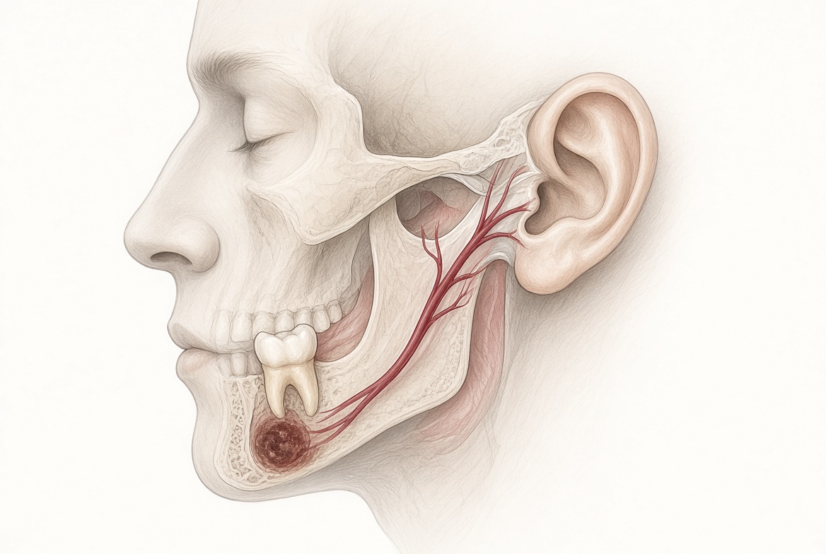
The neural mechanism is direct and anatomically well-established. The auriculotemporal nerve — a branch of the mandibular division of the trigeminal nerve (V3) — supplies sensory innervation to the skin of the ear, the external auditory canal, and the anterior wall of the tympanic membrane. It also innervates the temporomandibular joint and the posterior mandibular teeth. Nociceptive input from the dental pulp, the periapical tissues, or the periodontal ligament travels along V3 and can be referred — via the auriculotemporal nerve — to the ear as otalgia, even when the source is a tooth in the lower jaw with no local symptoms whatsoever.
The glossopharyngeal nerve (CN IX) provides an additional pathway: it innervates the posterior third of the tongue, the tonsillar region, and parts of the external ear canal. Pathology in the posterior oral cavity and oropharynx — including posterior dental infections — can generate ear pain through this second, independent neural route.
The Absence of Toothache Is Not Reassuring
Readers of earlier editions of this series will recognise this principle immediately. It applies here with equal and perhaps even greater force.
The patients who present with referred dental otalgia typically have no toothache. They have earache. They do not associate their ear symptoms with their teeth in any way, because their teeth do not hurt. Neither does their treating clinician — because the standard clinical question “do any of your teeth hurt?” receives a confident negative answer, and the dental hypothesis is abandoned before it was seriously considered.
This is the critical diagnostic error. As established throughout this series, occult periapical lesions — present in approximately one in four patients on CBCT — generate continuous trigeminal nociceptive input without producing conscious dental pain. That nociceptive input does not always manifest as jaw discomfort or facial pain. In some patients, the primary — or only — clinical expression of the subclinical dental nociception is ear pain, referred via the auriculotemporal branch of V3 to the external ear and tympanic membrane.
The patient experiences earache. The ear is normal. The tooth is silent. The CBCT finds the lesion. Treating the tooth resolves the ear pain.
This is the sequence we observe repeatedly in our practice — and it is supported by the pattern of referred otalgia aetiological studies: dental pathology is among the leading causes of ear pain that survives normal ENT examination and normal temporal bone imaging.
What We See in Our Practice
In our maxillofacial surgery practice in Pretoria and Benoni, we receive regular referrals for patients who have followed exactly the pathway described above. Multiple GP consultations. Repeated courses of antibiotics — for an infection that was never in the ear. ENT review with normal otoscopy and normal tympanometry. Medical CT of the temporal bone demonstrating no abnormality. The patient is frustrated, in pain, and without a diagnosis.
Our assessment protocol for these patients includes a structured clinical dental examination — specifically including pulp vitality testing on all posterior teeth on the symptomatic side — and full-mouth CBCT interpreted with attention to periapical pathology, periodontal bone loss, partially erupted or impacted teeth, and any bony or soft tissue abnormality.
In a significant proportion of these patients, CBCT identifies what conventional imaging and clinical examination missed: an occult periapical granuloma, failed endodontic treatment with persistent periapical infection, or advancing carious disease beneath an existing restoration. The tooth is asymptomatic. The patient has no toothache. But the lesion is present, confirmed on three-dimensional imaging, and generating the trigeminal nociceptive input that is being referred — via the auriculotemporal nerve — to the ear.
Following definitive dental treatment, the referred otalgia resolves. The antibiotics that were prescribed for months were treating an infection that was never in the ear canal. The true infection was in the jaw, hidden beneath the threshold of conventional imaging and beneath the threshold of conscious dental pain.
Why a Normal CT of the Temporal Bone Does Not Exclude a Dental Cause
When a patient reaches ENT and otalgia persists despite normal otoscopy, medical CT of the temporal bone is a reasonable and appropriate investigation to exclude middle ear, mastoid, and petrous bone pathology. It is the right tool for that question.
It is not, however, the right tool for detecting occult dental periapical pathology.
A medical CT of the temporal bone is calibrated and reconstructed for the middle ear structures. The dental root tips may be visible, but the scan is not optimised — in resolution, reconstruction plane, or field of view — for the detection of early-stage cortically-contained periapical granulomas in the dental alveolus. A negative temporal bone CT does not exclude a dental source of referred otalgia. It never could.
CBCT, calibrated with dental resolution, reconstructed in the orthogonal dental planes, and interpreted by a clinician specifically trained in maxillofacial and dental pathology, is the appropriate tool for this diagnostic question. It is the tool that closes the gap left by otoscopy, OPG, and temporal bone CT — detecting the occult dental lesion that each preceding investigation was structurally unable to find.
And the standard dental panoramic X-ray — if one was taken — is similarly insufficient. As established in every edition of this series, OPG misses approximately one in three periapical lesions detectable on CBCT, precisely because cortically-contained early lesions are structurally invisible on two-dimensional imaging.
A Word on Antibiotics
Antibiotic prescribing for persistent otalgia with a normal ear examination deserves specific comment.
Antibiotics are the appropriate first-line treatment for acute otitis media and otitis externa — when those conditions are confirmed on examination. When the ear examination is normal and no otological infection is identified, prescribing antibiotics treats nothing. There is no microbiological target. No bacterial infection has been demonstrated. The prescription is, in the most literal sense, a treatment for a disease the patient does not have.
The dental infection generating the referred otalgia — when present — is typically a chronic, contained periapical lesion. Even if periapical infection were theoretically accessible to systemic antibiotics, the necrotic root canal has no vascular supply. Adequate antibiotic concentrations cannot reach the intraradicular bacteria. Systemic antibiotics do not treat periapical lesions. Definitive treatment — root canal therapy or extraction — is required.
Repeated antibiotic prescribing for persistent otalgia with a normal ear delays the correct diagnosis, exposes the patient to antibiotic side effects and risks without benefit, and contributes to antimicrobial resistance. If the otoscope shows a normal ear, put down the prescription pad and think jaw.
The Diagnostic Pathway: A Structured Approach
For patients presenting with otalgia, the following structured approach is recommended:
- Step 1: Full clinical ear examination — otoscopy, tympanometry, pure tone audiometry. If positive findings are present, treat the identified otological condition. If the examination is normal, do not prescribe antibiotics. Proceed to Step 2.
- Step 2: With the ear assessed and found normal, check the jaw. Examine the temporomandibular joint — range of motion, joint tenderness, muscle palpation. Perform a comprehensive clinical dental examination including pulp vitality testing on all teeth in the symptomatic quadrant. Do not rely on the absence of dental pain as an exclusion criterion — the tooth will not hurt.
- Step 3: Full-mouth CBCT interpreted by a maxillofacial surgeon or oral radiologist, specifically to identify periapical pathology, failed endodontic treatment, and other occult dental or osseous pathology invisible on conventional 2D imaging.
- Step 4: Treat all identified dental pathology and review otalgia at four to six weeks. If the ear pain resolves, the dental source is confirmed.
- Step 5: If dental assessment is negative and otalgia persists, proceed to evaluation for other referred otalgia sources: cervical spine, tonsils and oropharynx, sinuses — and critically — head and neck malignancy, which can present as referred otalgia and must not be overlooked in patients with risk factors for malignant disease.
The key message: the ear and the jaw are examined together, not sequentially after years of failed treatment. With the ear, check the jaw. At the same consultation. At the first visit after a normal otoscopy.
The Broader Picture: Four Editions, One Principle
This series has now traced the same clinical argument across four distinct diagnostic scenarios:
- A hip or knee replacement patient develops a catastrophic prosthetic joint infection from a silent dental lesion
- A patient spends years in failed TMJ treatment while a tooth silently drives their bruxism and joint loading
- A patient is diagnosed with trigeminal neuralgia and placed on powerful neurological medication when a tooth is the true source of their facial pain
- A patient receives repeated antibiotics and normal ENT investigations for earache that originates not in the ear, but in the jaw
In every case: the pathology is common. It is asymptomatic. It is missed by the imaging tools used in the standard diagnostic pathway. CBCT detects it. Treating it resolves the problem that no other intervention could fix.
Before the joint, check the jaw.
Before the nerve, check the jaw.
With the ear, check the jaw.
The jaw is not an afterthought. It is not the last referral after everything else has failed. It is a first-line diagnostic consideration — in otalgia, in facial pain, in jaw joint dysfunction, and in pre-operative surgical assessment.
For referrals or further information about CBCT-based dental assessment for referred otalgia, facial pain, TMJ dysfunction, or pre-operative arthroplasty screening, contact our rooms at:
Dr. Frits Hoogendijk | Maxillofacial Surgeon
Menlopark Medical Centre, Pretoria | Benoni
082 390 7425
This article is intended for general information purposes. Clinical decisions should always be made in consultation with qualified healthcare professionals based on individual patient assessment.
References
- Charlett SD, Coatesworth AP. Referred otalgia: a structured approach to diagnosis and treatment. Int J Clin Pract. 2007;61(6):1015–1021.
- Chen RC, et al. Referred otalgia: common causes and evidence-based strategies for assessment and management. Can Fam Physician. 2023.
- Kim DS, et al. A study of the etiology of referred otalgia. Korean J Audiol. 2013.
- Khan MA, et al. The frequency of involvement of head and neck sites in referred otalgia — an experience at a tertiary care hospital. PMC. 2019.
- Klasser GD, de Leeuw R. Orofacial Pain: Guidelines for Assessment, Diagnosis and Management. 7th ed. Quintessence; 2023.
- Kanagasingam S, et al. Diagnostic accuracy of periapical radiography and cone beam computed tomography in detecting apical periodontitis. Int Endod J. 2017;50(5):417–426.
- Bornstein MM, et al. Cone beam computed tomography in endodontics: a systematic review. J Endod. 2015;41(10):1591–1597.
- Lofthag-Hansen S, et al. Limited cone-beam CT and intraoral radiography for the diagnosis of periapical pathology. Oral Surg Oral Med Oral Pathol Oral Radiol Endod. 2007;103(1):114–119.
- Abbott PV. Classification, diagnosis and clinical manifestations of apical periodontitis. Endodontic Topics. 2004;8(1):36–54.
- Okeson JP. Management of Temporomandibular Disorders and Occlusion. 8th ed. Elsevier; 2019.
- International Headache Society. ICHD-3. Cephalalgia. 2018;38(1):1–211.
- Sander T, et al. PMC. The interaction between the nervous system and the stomatognathic system. PMC. 2023. PMC10425412.
- Luscan R, et al. TMJ arthritis is a frequent complication of otomastoiditis. Int J Pediatr Otorhinolaryngol. 2016.
- Semantic Scholar. Review article: TMD as a cause of referred otalgia and differential diagnosis for ENT clinicians.
- Royal College of Surgeons England. Faculty of Dental Surgery Clinical Guidelines: Diagnosis and Management of Disorders of the Temporomandibular Joint. RCS England; 2013.