BEFORE THE JOINT, CHECK THE JAW: EDITION TWO
When Your Jaw Joint Is Not the Problem — It Is the Victim
By Dr. Frits Hoogendijk | Maxillofacial Surgeon | Menlopark Medical Centre, Pretoria
Contact: 082 390 7425
In our first article in this series, we explored why every patient scheduled for a hip or knee replacement should undergo a cone beam CT scan of the jaw before surgery, and why silent tooth infections are a preventable cause of catastrophic prosthetic joint infection.
This second edition takes the same principle into a different but equally important clinical territory: the temporomandibular joint, or TMJ. If you have been told you have temporomandibular disorder (TMD), or if you are a clinician treating patients with jaw pain, clicking, limited opening, or chronic headaches, this article raises a question that is rarely asked early enough:
Before we treat the jaw joint, have we checked whether the jaw joint is actually the problem?
What Is the Temporomandibular Joint — and What Is TMD?
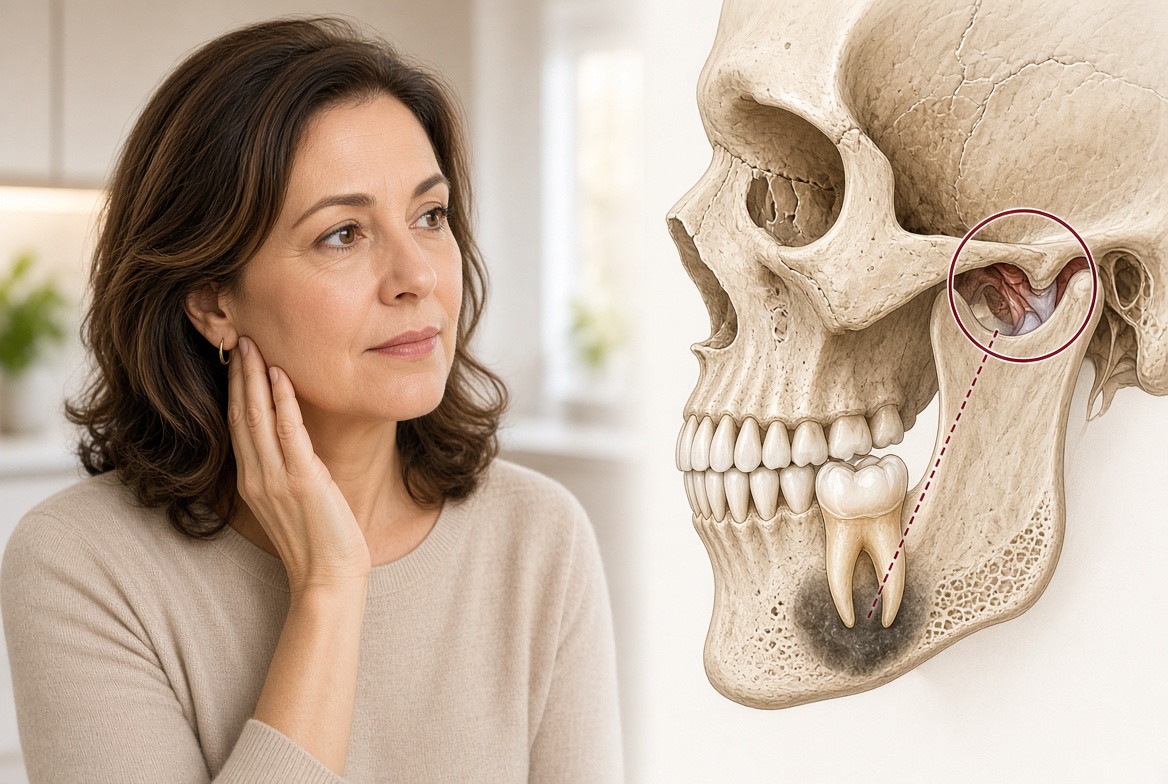
The temporomandibular joint connects the lower jaw to the base of the skull, just in front of each ear. It is one of the most complex joints in the body, capable of hinging, gliding and rotating, and active thousands of times a day in chewing, speaking, swallowing and yawning.
Temporomandibular disorder is an umbrella term for conditions affecting this joint and the muscles that control jaw movement. Symptoms include pain in front of the ear, clicking or popping, difficulty opening fully, jaw locking, temple headaches, and pain in the cheek or jaw muscles.
TMD is common. It affects an estimated 5–12% of the population to some degree, and it is the second most frequent cause of facial pain after toothache. Treatment ranges from splints, physiotherapy and anti-inflammatory medication to joint injections, arthrocentesis, and in severe cases surgery.
A significant proportion of patients who receive a TMD diagnosis, and who undergo treatment directed at the joint, are not primarily suffering from joint pathology. The joint has been damaged. The damage was caused by something else: a tooth.
The Tooth That Did Not Hurt — But Still Caused the Problem
This is the most important concept in this article, and the one most frequently misunderstood.
The absence of toothache is not reassuring. It is irrelevant.
The dental pathology most responsible for driving jaw-joint dysfunction is, by definition, the pathology that produces no conscious dental pain. A tooth that hurts gets attended to. The dangerous tooth is the one the patient cannot feel.
A periapical granuloma, a chronic infection at the root tip, generates nociceptive signals through the trigeminal nerve continuously, below the threshold of conscious awareness. The patient has no toothache, no sensitivity to hot or cold, and the tooth feels normal to tapping. Inside the jawbone, an established infection is firing low-level signals into the trigeminal system, modulating masticatory muscle activity, increasing bruxism, and loading the joint, every day and every night.
This is neuroscience, not theory. The trigeminal nucleus receives continuous afferent input from the periapical tissues. That input does not need to reach the cortex as conscious pain to alter motor output to the jaw muscles. Subcortical pain-adaptation reflexes increase masseter and temporalis tone in response to nociceptive dental input, even during sleep.
Asking a patient whether their teeth hurt tells you almost nothing about whether a tooth is driving their TMJ dysfunction. The answer will almost always be no. That is precisely the problem.
What We See in Our Practice
In our maxillofacial practice we regularly receive referrals for patients who have undergone prolonged treatment for TMJ dysfunction: splints for months or years, repeated physiotherapy, joint injections, arthrocentesis, without sustained improvement. The joint pathology was real. The treatment was appropriate for the joint. It was not succeeding because the joint was not the origin of the problem.
In a number of these patients, CBCT identifies an occult periapical lesion, a chronic granuloma or cyst, invisible on the panoramic X-rays taken during dental assessment. The tooth is asymptomatic, often root-treated or heavily restored, and assumed to be a non-issue. Following extraction, masticatory muscle activity settles, bruxism reduces, and TMJ symptoms that had resisted years of joint-directed treatment resolve or substantially improve.
The pattern is consistent: failed TMD treatment, occult dental lesion on CBCT, tooth removed, joint settles. Remove the nociceptive driver, and the motor cascade that was loading the joint is extinguished. The treatment directed at the joint was not wrong. It was incomplete, because the source was never found.
The Invisible Tooth: Pathologies That Drive the Cascade
Beyond the obvious impacted third molar, the lesions most likely to go undetected include:
- Silent periapical granulomas and cysts. Cortically contained chronic infections, present in about one in four patients on CBCT, and invisible on panoramic X-ray in about one in three cases. Continuously generating trigeminal input.
- Advanced decay beneath restorations. Caries under a filling or crown, invisible clinically and often obscured by restoration scatter. Early pulpitis can generate nociceptive input below the threshold of conscious pain.
- Failed root canal therapy. Root-treated teeth are widely assumed to be inert. Persistent periapical infection is common, and the tooth typically remains asymptomatic. Conventional radiography is particularly unreliable here. CBCT is the most sensitive available tool.
- Cracked tooth syndrome. An incomplete fracture of a posterior tooth produces sharp, unpredictable pain on specific loading, often on release. The patient shifts the bite to protect the tooth, creating eccentric jaw posture and asymmetric condylar loading. The crack is frequently invisible on conventional radiography.
Each can produce the sustained subclinical trigeminal nociception that drives muscle hyperactivity, bruxism and secondary TMJ loading. None reliably produces toothache. All are detectable on CBCT.
Why Standard Dental X-Rays Are Not Sufficient
Conventional two-dimensional radiography, periapical films and panoramic OPG, requires 30–50% of the surrounding cortical bone to have been destroyed before a periapical lesion becomes detectable. Early lesions confined to cancellous bone are structurally invisible.
- CBCT sensitivity for periapical lesion detection: 89–96%
- Periapical radiograph sensitivity: 38–77%
- Panoramic OPG sensitivity: 33–82%
Studies comparing the two modalities in the same patients have found that CBCT reveals up to 2.5 times more periapical pathology than digital periapical radiography. Approximately one in three CBCT-detectable lesions is completely invisible on panoramic imaging.
A dental clearance based on a panoramic X-ray and clinical examination, in a patient with no dental complaints, may provide false reassurance. The Royal College of Surgeons of England guideline on painful temporomandibular disorders states that imaging of the dentulous regions should form part of investigation to identify sources of odontogenic or referred pain which may mimic TMD. Those recommendations have not been turned into a systematic protocol, and conventional imaging is the wrong tool for the job.
What Happens When You Treat the Joint Without Treating the Tooth
The patient receives an occlusal splint. It provides some relief, but the nociceptive driver continues. Physiotherapy improves mobility, then muscle hyperactivity returns. Joint injections or arthrocentesis calm the joint for a period, then symptoms recur. The patient is labelled difficult or treatment-resistant. More invasive options are considered. The clinician, operating in good faith, escalates treatment in a direction that cannot succeed, because the driving force is still present.
All of this is preventable. In many of these patients the solution is a single CBCT, an occult lesion identified, and an extraction.
The Proposed Approach: Check the Jaw First
Before initiating or escalating TMJ-directed treatment, we recommend this sequence:
- A comprehensive clinical dental examination, including pulp vitality testing on all posterior teeth, assessment for crack sensitivity, and periodontal evaluation, regardless of whether the patient reports dental pain.
- A full-mouth CBCT, interpreted by a maxillofacial surgeon with specific training in jaw and dental pathology.
- Identify and treat all occult dental pathology: periapical lesions, failed root canal therapy, advancing caries, cracked teeth.
- Allow four to six weeks after dental treatment for masticatory muscle adaptation and reduction in bruxism.
- Reassess TMJ symptoms. If they have resolved or substantially reduced, the dental pathology was the primary driver.
- If symptoms persist after elimination of dental nociceptive sources, proceed with confidence that the TMJ is the primary pathology, and that joint-directed treatment will not be undermined by an ongoing dental driver.
The sequence is straightforward, low-risk and low-radiation. Its potential benefit is avoiding months or years of treatment failure in patients whose joint dysfunction was driven by a tooth that no one looked for carefully enough.
The Broader Principle
Both editions of this series rest on the same clinical philosophy. Joints, whether prosthetic or native, in the hip, the knee or the jaw, are targets. They present with real pathology. The origin of that pathology is not always the joint itself.
The jaw is the first link in the chain. Before the joint, check the jaw.
In prosthetic joint replacement: check for occult periapical lesions that could drive haematogenous infection of the new implant. In temporomandibular dysfunction: check for occult dental nociceptive drivers loading the joint through muscular hyperactivity and bruxism. In both contexts the tool is the same, and the cost of not looking is far greater than the cost of a CBCT scan.
For referrals or further information about pre-treatment CBCT assessment for TMD, or pre-operative oral screening for arthroplasty, contact the rooms:
Dr Frits Hoogendijk | Maxillofacial Surgeon
Suite 4, Menlopark Medical Centre, Pretoria
Room 107, Lakeview Hospital, 1 Mowbray Avenue, Benoni
082 390 7425 | 082 855 2389 | admin@theface.co.za
This article is intended for general information. Clinical decisions should be made in consultation with a qualified healthcare professional, based on individual assessment.
References
- Abbott PV. Classification, diagnosis and clinical manifestations of apical periodontitis. Endodontic Topics. 2004;8(1):36–54.
- American Academy of Orofacial Pain. Orofacial Pain: Guidelines for Assessment, Diagnosis and Management. 6th ed. Quintessence; 2018.
- Kanagasingam S, et al. Diagnostic accuracy of periapical radiography and cone beam computed tomography in detecting apical periodontitis using histopathology as reference standard. Int Endod J. 2017;50(5):417–426.
- De Leeuw R, Klasser GD, eds. Orofacial Pain: Guidelines for Assessment, Diagnosis and Management. American Academy of Orofacial Pain; 2018.
- Slade GD, Ohrbach R, Greenspan JD, et al. Painful temporomandibular disorder: decade of discovery from OPPERA studies. J Dent Res. 2016;95(10):1084–1092.
- Nixdorf DR, Moana-Filho EJ, Law AS, et al. Frequency of persistent tooth pain after root canal therapy: a systematic review and meta-analysis. J Endod. 2010;36(2):224–230.
- Türp JC, Komine F, Hugger A. Efficacy of stabilization splints for the management of patients with masticatory muscle pain: a qualitative systematic review. Clin Oral Investig. 2004;8(4):179–195.
- Peck CC, Goulet JP, Lobbezoo F, et al. Expanding the taxonomy of the diagnostic criteria for temporomandibular disorders (DC/TMD). J Oral Rehabil. 2014;41(1):2–23.
- American Academy of Family Physicians. Temporomandibular disorders: differential diagnosis and management. Am Fam Physician. 2015;91(6):378–386.
- Lobbezoo F, Ahlberg J, Glaros AG, et al. Bruxism defined and graded: an international consensus. J Oral Rehabil. 2013;40(1):2–4.
- Lavigne GJ, Khoury S, Abe S, Yamaguchi T, Raphael K. Bruxism physiology and pathology: an overview for clinicians. J Oral Rehabil. 2008;35(7):476–494.
- Manfredini D, Lobbezoo F. Relationship between bruxism and temporomandibular disorders: a systematic review of literature from 1998 to 2008. Oral Surg Oral Med Oral Pathol Oral Radiol Endod. 2010;109(6):e26–e50.
- Bornstein MM, et al. Cone beam computed tomography in endodontics: a systematic review of its efficacy to diagnose periapical pathology. J Endod. 2015;41(10):1591–1597.
- Manfredini D, Guarda-Nardini L, Winocur E, et al. Research diagnostic criteria for temporomandibular disorders: a systematic review of axis I epidemiologic findings. Oral Surg Oral Med Oral Pathol Oral Radiol Endod. 2011;112(4):453–462.
- Comparative analysis: CBCT versus conventional radiography for periapical lesion detection. J Endod. 2023;49(3):247–254.
- Debelian GJ, Olsen I, Tronstad L. Bacteremia in conjunction with endodontic therapy. Endod Dent Traumatol. 1995;11(3):142–149.
- Okeson JP. Management of Temporomandibular Disorders and Occlusion. 8th ed. Elsevier; 2019.
- Huang GJ, LeResche L, Critchlow CW, Martin MD, Drangsholt MT. Risk factors for diagnostic subgroups of painful temporomandibular disorders. J Dent Res. 2002;81(4):284–288.
- Michelotti A, Iodice G. The role of orthodontics in temporomandibular disorders. J Oral Rehabil. 2010;37(6):411–429.
- Roberts GJ. Dentists are innocent! “Everyday” bacteremia is the real culprit. Pediatr Cardiol. 1999;20(5):317–325.
- Manfredini D, et al. Bruxism and temporomandibular disorders: a clinical comparison. Clin Oral Investig. 2015;19(9):2141–2148.
- Lofthag-Hansen S, et al. Limited cone-beam CT and intraoral radiography for the diagnosis of periapical pathology. Oral Surg Oral Med Oral Pathol Oral Radiol Endod. 2007;103(1):114–119.
- Lobbezoo F, Ahlberg J, Raphael KG, et al. International consensus on the assessment of bruxism: report of a work in progress. J Oral Rehabil. 2018;45(11):837–844.
- European Society of Endodontology (ESE). Quality guidelines for endodontic treatment: consensus report. Int Endod J. 2006;39(12):921–930.
- Lavigne GJ, Huynh N, Kato T, et al. Genesis of sleep bruxism: motor and autonomic-cardiac interactions. Arch Oral Biol. 2007;52(4):381–384.
- Lund JP, Donga R, Widmer CG, Stohler CS. The pain-adaptation model: a discussion of the relationship between chronic musculoskeletal pain and motor activity. Can J Physiol Pharmacol. 1991;69(5):683–694.
- Nixdorf DR, Law AS, Look JO, et al. Large-scale clinical endodontic research in the National Dental Practice-Based Research Network: study overview and methods. J Endod. 2012;38(11):1470–1478.
- Molander A, et al. The relevance of orthograde retreatment in root-filled teeth with periapical lesions: panoramic vs periapical imaging. Oral Surg Oral Med Oral Pathol Oral Radiol. 2015;120(6):786–792.
- Svensson P, Jadidi F, Arima T, Arendt-Nielsen L, Farina D. Relationships between craniofacial pain and bruxism. J Oral Rehabil. 2008;35(7):524–547.
- Lund JP, Sessle BJ. Neurophysiological mechanisms in temporomandibular pain and dysfunction. In: Sessle BJ, Bryant PS, Dionne RA, eds. Temporomandibular Disorders and Related Pain Conditions. IASP Press; 1995.
- Stohler CS. Masticatory myalgias: emphasis on the nerve growth factor-estrogen link. Pain Forum. 1997;6(3):176–180.
- Dworkin SF, LeResche L. Research diagnostic criteria for temporomandibular disorders: review, criteria, examinations and specifications, critique. J Craniomandib Disord. 1992;6(4):301–355.
- Poveda-Roda R, Bagán JV, Díaz-Fernández JM, Hernández-Bazán S, Jiménez-Soriano Y. Review of temporomandibular joint pathology. Part I: classification, epidemiology and risk factors. Med Oral Patol Oral Cir Bucal. 2007;12(4):E292–298.
- Shaffer SM, Brismée JM, Sizer PS, Courtney CA. Temporomandibular disorders. Part 1: anatomy and examination/diagnosis. J Man Manip Ther. 2014;22(1):2–12.
- Truelove E, Pan W, Look JO, et al. The research diagnostic criteria for temporomandibular disorders. III: validity of Axis I diagnoses. J Orofac Pain. 2010;24(1):35–47.
- Imhoff B, Ahlers MO. Cracked tooth syndrome — diagnostics and therapy. Quintessence Int. 2020;51(2):100–110.
- Tanaka E, Detamore MS, Mercuri LG. Degenerative disorders of the temporomandibular joint: etiology, diagnosis, and treatment. J Dent Res. 2008;87(4):296–307.
- Owatz CB, Khan AA, Schindler WG, Schwartz SA, Keiser K, Hargreaves KM. The incidence of mechanical allodynia in patients with irreversible pulpitis. J Endod. 2007;33(5):552–556.
- Sessle BJ. Acute and chronic craniofacial pain: brainstem mechanisms of nociceptive transmission and neuroplasticity, and their clinical correlates. Crit Rev Oral Biol Med. 2000;11(1):57–91.
- American Association of Oral and Maxillofacial Surgeons. Evidence-Based Recommendations for the Use of CBCT in Oral and Maxillofacial Surgery. AAOMS; 2022.
- Royal College of Surgeons England. Faculty of Dental Surgery Clinical Guidelines: Diagnosis and Management of Disorders of the Temporomandibular Joint. RCS England; 2013.